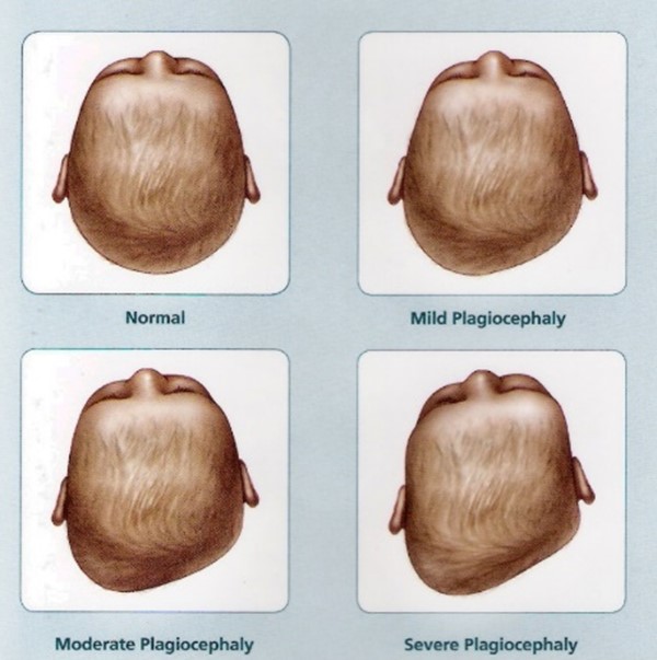
Positional Plagiocephaly is a common condition in infants characterized by asymmetrical flattening at the back of the head as a result of external mechanical pressure.
Plagiocephaly is identified in 33%-47% of infants within the first year of life. Growing evidence shows that Plagiocephaly can lead to permanent deformation of the head, neck and face, and may have developmental implications including learning disabilities, language disorders, perceptual & sensorimotor problems, and poorer social skills. However, this condition is treatable through correcting physical interventions or helmets.
Clinical detection of plagiocephaly in infants relies on observational diagnostics using a scale such as the Argenta classification that classifies plagiocephaly into five degrees of severity according to specific cranial asymmetry characteristics.
This study integrated a clinical aim followed by an algorithmic one. First we examined the reliability of the clinical Argenta classification and managed to show it is reliable and applicable for clinical use. We then developed an automated system for objectively evaluating severity of plagiocephaly in infants. This allows an objective, reproducible and comparable evaluation of the severity of the condition. It will enable quantitative evaluation of effects of treatment as well as fair comparison across patients.
We developed a method to predict Argenta scores from three 2D views of the infant’s head.

First, testing was performed to ensure that the Argenta score can indeed be assessed from these three views and that the score is reliable and validated (paper in preparation).
Using Image Processing and Computer Vision methods, we extract features from the images and use them to predict the Argenta scores. The computerized approach follows the physiotherapist methodology of plagiocephaly assessment, such as using a virtual caliper to measure diameter of head, measuring displacement of the ears, asymmetry of the face, flattening of the skull and more.

This study was performed by an interdisciplinary team: Dr Hilla Sarig-Bahat from the Dept of Physical Therapy, Head of Orthopedic Division and Prof. Hel-Or from the Dept of Computer Science and the Computational Human Behavior Lab. This study was supported by the University of Haifa’s Data Science Research Center.
Positional Plagiocephaly is a common condition in infants characterized by asymmetrical flattening at the back of the head as a result of external mechanical pressure.
Plagiocephaly is identified in 33%-47% of infants within the first year of life. Growing evidence shows that Plagiocephaly can lead to permanent deformation of the head, neck and face, and may have developmental implications including learning disabilities, language disorders, perceptual & sensorimotor problems, and poorer social skills. However, this condition is treatable through correcting physical interventions or helmets.
Clinical detection of plagiocephaly in infants relies on observational diagnostics using a scale such as the Argenta classification that classifies plagiocephaly into five degrees of severity according to specific cranial asymmetry characteristics.
This study integrated a clinical aim followed by an algorithmic one. First we examined the reliability of the clinical Argenta classification and managed to show it is reliable and applicable for clinical use. We then developed an automated system for objectively evaluating severity of plagiocephaly in infants. This allows an objective, reproducible and comparable evaluation of the severity of the condition. It will enable quantitative evaluation of effects of treatment as well as fair comparison across patients.
We developed a method to predict Argenta scores from three 2D views of the infant’s head.
First, testing was performed to ensure that the Argenta score can indeed be assessed from these three views and that the score is reliable and validated (paper in preparation).
Using Image Processing and Computer Vision methods, we extract features from the images and use them to predict the Argenta scores. The computerized approach follows the physiotherapist methodology of plagiocephaly assessment, such as using a virtual caliper to measure diameter of head, measuring displacement of the ears, asymmetry of the face, flattening of the skull and more.
This study was performed by an interdisciplinary team: Dr Hilla Sarig-Bahat from the Dept of Physical Therapy, Head of Orthopedic Division and Prof. Hel-Or from the Dept of Computer Science and the Computational Human Behavior Lab. This study was supported by the University of Haifa’s Data Science Research Center.